Dinosaur Aunts, Bacterial Stowaways, & Insect Milk
Milk is everywhere. From the dairy aisle at the grocery store to the explosive cover of the Mother’s Day issue of Time magazine, the ubiquity of milk makes it easy to take for granted. But surprisingly, milk synthesis is evolutionarily older than mammals. Milk is even older than dinosaurs. Moreover, milk contains constituents that infants don’t digest, namely oligosaccharides, which are the preferred diet of the neonate’s intestinal bacteria (nom nom nom!) And milk doesn’t just feed the infant, and the infant’s microbiome; the symbiotic bacteria are IN mother’s milk.
The fossil record, unfortunately, leaves little direct evidence of the soft-tissue structures that first secreted milk. Despite this, paleontologists can scrutinize morphological features of fossils, such as the presence or absence of milk teeth (diphyodonty), to infer clues about the emergence of “milk.” Genome-wide surveys of the expression and function of mammary genes across divergent taxa, and experimental evo-devo manipulations of particular genes also yield critical insights. As scientists begin to integrate information from complementary approaches, a clearer understanding of the evolution of lactation emerges.

In his recent paper, leading lactation theorist Dr. Olav Oftedal discusses the ancient origins of milk secretion (2012). He contends the first milk secretions originated ~310 million years ago (MYA) in synapsids, a lineage ancestral to mammals and contemporaries with sauropsids, the ancestors of reptiles, birds, and dinosaurs. Synapsids and sauropsids produced eggs with multiple membrane layers, known as amniote eggs. Such eggs could be laid on land. However, synapsid eggs had permeable, parchment-like shells and were vulnerable to water loss. Burying these eggs in damp soil or sand near water resources- like sea turtles do- wasn’t an option, posits Oftedal. The buried temperatures would have likely been too cold for the higher metabolism of synapsids. But incubating eggs in a nest would have evaporated water from the egg. The synapsid egg was proverbially between a rock and a hard place: too warm to bury, too permeable to incubate.

Luckily for us, a mutation gave rise to secretions from glandular skin on the belly of the synapsid parent. This mechanism replenished water lost during incubation, allowing synapsids to lay eggs in a variety of terrestrial environments. As other mutations randomly arose and were favored by selection, milk composition became increasingly complex, incorporating nutritive, protective, and hormonal factors (Oftedal 2012). Some of these milk constituents are shunted into milk from maternal blood, some- although also present in the maternal blood stream- are regulated locally in the mammary gland, and some very special constituents are unique to milk. Lactose and oligosaccharides (a sugar with lactose at the reducing end) are two constituents unique to mammalian milk, but are interestingly divergent among mammals living today.


Marsupial and monotreme young seemingly digest oligosaccharides. Among placental mammals, however, young do not have the requisite enzymes in their stomach and small intestine to utilize oligosaccharides themselves. In humans, oligosaccharides are the 4th main constituent of milk (after water, lactose, and fat). Why do eutherian mothers synthesize oligosaccharides in milk, if infants don’t digest them?
In May, Anna Petherick’s post “Multi-tasking Milk Oligosaccharides” revealed that oligosaccharides serve a number of critical roles for supporting the healthy colonization and maintenance of the infant’s intestinal microbiome. Beneficial bacterial symbionts contribute to the digestion of nutrients from our food. Just as importantly, they are an essential component of the immune system, defending their host against many ingested pathogens. The structures of milk oligosaccharides have been described for a number of primates, including humans, and data are now available from all major primate clades; strepsirrhines (i.e. lemurs), New World monkey (i.e. capuchin), Old World monkey (i.e. rhesus), and apes (i.e. chimpanzee).

Among all non-human primates studied to date, Type II oligosaccharides are most prevalent (Type II oligosaccharides contain lacto-N-biose I). Type I oligosaccharides (containing N-acetyllactosamine) are absent, or in much lower concentrations than Type II (Taufik et al. 2012).
In human milk, there is a much greater diversity and higher abundance of milk oligosaccharides than found in the milk of other primates. Most primate taxa have between 5-30 milk oligosaccharides; humans have ~200. Even more astonishingly, humans predominantly produce Type I oligosaccharides, the preferred food of the most prevalent bacterium in the healthy human infant gut- Bifidobacteria(Urashima et al 2012, Taufik et al. 2012).

Human infants have bigger brains and an earlier age at weaning than do our closest ape relatives. Many anthropologists have hypothesized that constituents in mother’s milk, such as higher fat concentrations or unique fatty acids, underlie these differences in human development. But only oligosaccharides, a constituent that the human infant does not itself utilize, are demonstrably derived from our primate relatives (Hinde and Milligan 2011). At some point in human evolution there must have been strong selective pressure to optimize the symbiotic relationship between the infant microbiome and the milk mothers synthesize to support it. The human and Bifidobacteria genomes show signatures of co-evolution, but the selective pressures and their timing remain to be understood (Martin and Sela 2013).


Evolutionary Origins of Lactation
The fossil record, unfortunately, leaves little direct evidence of the soft-tissue structures that first secreted milk. Despite this, paleontologists can scrutinize morphological features of fossils, such as the presence or absence of milk teeth (diphyodonty), to infer clues about the emergence of “milk.” Genome-wide surveys of the expression and function of mammary genes across divergent taxa, and experimental evo-devo manipulations of particular genes also yield critical insights. As scientists begin to integrate information from complementary approaches, a clearer understanding of the evolution of lactation emerges.

In his recent paper, leading lactation theorist Dr. Olav Oftedal discusses the ancient origins of milk secretion (2012). He contends the first milk secretions originated ~310 million years ago (MYA) in synapsids, a lineage ancestral to mammals and contemporaries with sauropsids, the ancestors of reptiles, birds, and dinosaurs. Synapsids and sauropsids produced eggs with multiple membrane layers, known as amniote eggs. Such eggs could be laid on land. However, synapsid eggs had permeable, parchment-like shells and were vulnerable to water loss. Burying these eggs in damp soil or sand near water resources- like sea turtles do- wasn’t an option, posits Oftedal. The buried temperatures would have likely been too cold for the higher metabolism of synapsids. But incubating eggs in a nest would have evaporated water from the egg. The synapsid egg was proverbially between a rock and a hard place: too warm to bury, too permeable to incubate.
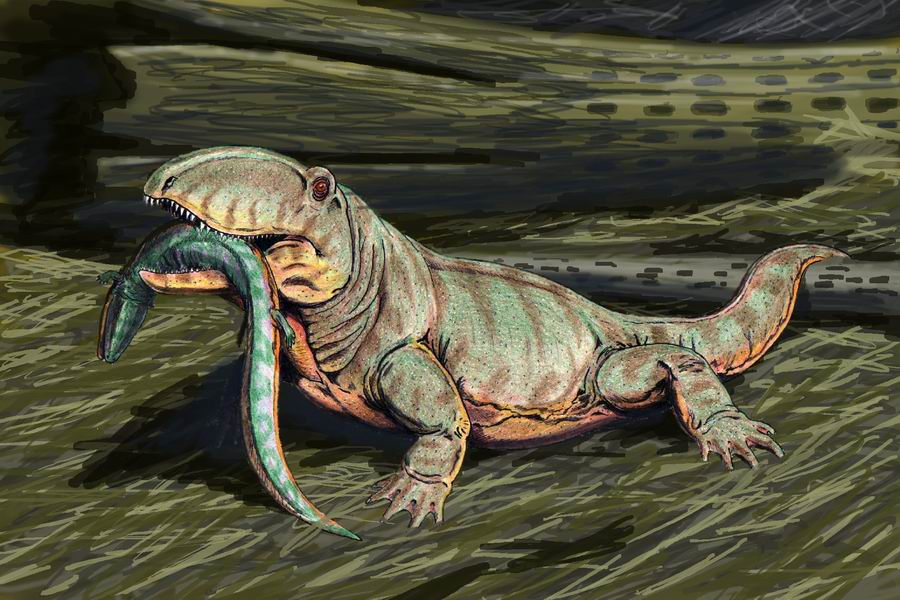
Ophiacodon by Dmitri Bogdanov
Luckily for us, a mutation gave rise to secretions from glandular skin on the belly of the synapsid parent. This mechanism replenished water lost during incubation, allowing synapsids to lay eggs in a variety of terrestrial environments. As other mutations randomly arose and were favored by selection, milk composition became increasingly complex, incorporating nutritive, protective, and hormonal factors (Oftedal 2012). Some of these milk constituents are shunted into milk from maternal blood, some- although also present in the maternal blood stream- are regulated locally in the mammary gland, and some very special constituents are unique to milk. Lactose and oligosaccharides (a sugar with lactose at the reducing end) are two constituents unique to mammalian milk, but are interestingly divergent among mammals living today.
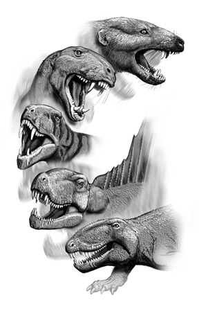
Illustration by Carl Buell
Mammalian and Primate Divergences: Milk Composition
Among all mammals studied to date, lactose and oligosaccharides are the primary sugars in milk. Lactose is synthesized in mammary glands only. Urashima and colleagues explain that lactose synthesis is contingent on the mammalian-specific protein alpha-lactalbumin (2012). Alpha-lactalbumin is very similar in amino-acid structure to C-type lysozyme, a more ancient protein found throughout vertebrates and insects. C-type lysozyme acts as an anti-bacterial agent. Oligosaccharides are predominant in the milks of marsupials and egg-laying monotremes (i.e. the platypus), but lactose is the most prevalent sugar in the milk of most placental (aka eutherian) mammals. Interestingly, the oligosaccharides in the milk of placental mammals are most similar to the oligosaccharides in the milk of monotremes. Unique oligosaccharides in marsupial milk emerged after the divergence of placental mammals.
Marsupial and monotreme young seemingly digest oligosaccharides. Among placental mammals, however, young do not have the requisite enzymes in their stomach and small intestine to utilize oligosaccharides themselves. In humans, oligosaccharides are the 4th main constituent of milk (after water, lactose, and fat). Why do eutherian mothers synthesize oligosaccharides in milk, if infants don’t digest them?
In May, Anna Petherick’s post “Multi-tasking Milk Oligosaccharides” revealed that oligosaccharides serve a number of critical roles for supporting the healthy colonization and maintenance of the infant’s intestinal microbiome. Beneficial bacterial symbionts contribute to the digestion of nutrients from our food. Just as importantly, they are an essential component of the immune system, defending their host against many ingested pathogens. The structures of milk oligosaccharides have been described for a number of primates, including humans, and data are now available from all major primate clades; strepsirrhines (i.e. lemurs), New World monkey (i.e. capuchin), Old World monkey (i.e. rhesus), and apes (i.e. chimpanzee).

Among all non-human primates studied to date, Type II oligosaccharides are most prevalent (Type II oligosaccharides contain lacto-N-biose I). Type I oligosaccharides (containing N-acetyllactosamine) are absent, or in much lower concentrations than Type II (Taufik et al. 2012).
In human milk, there is a much greater diversity and higher abundance of milk oligosaccharides than found in the milk of other primates. Most primate taxa have between 5-30 milk oligosaccharides; humans have ~200. Even more astonishingly, humans predominantly produce Type I oligosaccharides, the preferred food of the most prevalent bacterium in the healthy human infant gut- Bifidobacteria(Urashima et al 2012, Taufik et al. 2012).
Vertical Transmission of Bacteria via Milk
In the womb, the infant is largely protected from maternal bacteria due to the placental barrier. But upon birth, the infant is confronted by a teeming microbial milieu that is both a challenge and an opportunity. The first inoculation of commensal bacteria occurs during delivery as the infant passes through the birth canal and is exposed to a broad array of maternal microbes. Infants born via C-section are instead, and unfortunately, colonized by the microbes “running around” the hospital. But exposure to the mother’s microbiome continues long after birth. Evidence for vertical transmission of maternal bacteria via milk has been shown in rodents, monkeys(Jin et al. 2011), humans(Martin et al. 2012), and… insects.
Yes, INSECTS!
Yes, INSECTS!
A number of insects have evolved the ability to rely on nutritionally incomplete food sources. They are able to do so because bacteria that live inside their cells provide what the food does not. These bacteria are known as endosymbionts and the specialized cells the host provides for them to live in are called bacteriocytes. For example, the tsetse fly has a bacterium, Wigglesworthia glossinidia,* that provides B vitamins not available from blood meals. Um, if you are squeamish, don’t read the previous sentence.
*I submit the tsetse fly and its bacterial symbiont (Wigglesworthia glossinidia)
for consideration as the number one mutualism in which the common name of the host
and the Latin name of the bacteria are awesome to say out loud!
Bring on your challenger teams.
for consideration as the number one mutualism in which the common name of the host
and the Latin name of the bacteria are awesome to say out loud!
Bring on your challenger teams.
Hosokawa and colleagues recently revealed the Russian nesting dolls that are bats (Miniopterus fuliginosus), bat flies (Nycteribiidae), and endosymbiotic bacteria (proposed name Aschnera chenzii)(2012). Bat flies are the obligate ectoparasites of bats (Peterson et al. 2007). They feed on the blood of their bat hosts, and for nearly their entire lifespan, bat flies live in the fur of their bat hosts. Females briefly leave their host to deposit pupae on stationary surfaces within the bat roost.
 Bat flies are even more crazy amazing because they have a uterus and provide MILK internally through the uterus to larva! Male and female bat flies have endosymbiotic bacteria living in bacteriocytes along the sides of their abdominal segments (revealed by 16S rRNA). Additionally, females host bacteria inside the milk gland tubules, “indicating the presence of endosymbiont cells in milk gland secretion”.
The authors are not yet certain of the specific nutritional role that these bacterial endosymbionts play in the bat fly host. The bacteria may provide B vitamins, as other bacterial symbionts of blood-consuming insects are known to do. My main question is what is the exact role of the bacteria in the milk gland tubules? Are they there to add nutritional value to the milk for the larva, to stowaway in milk for vertical transmission to larva, or both?

The authors are not yet certain of the specific nutritional role that these bacterial endosymbionts play in the bat fly host. The bacteria may provide B vitamins, as other bacterial symbionts of blood-consuming insects are known to do. My main question is what is the exact role of the bacteria in the milk gland tubules? Are they there to add nutritional value to the milk for the larva, to stowaway in milk for vertical transmission to larva, or both?
Ernst Haeckel Chiroptera
Conclusions
The studies described above represent new frontiers in lactation research. The capacity to secrete “milk” has been evolving since before the age of dinosaurs, but we still know relatively little about the diversity of milks produced by mammals today. Even less understood are the consequences and functions of various milk constituents in the developing neonate. Despite the many unknowns, it is increasingly evident that mother’s milk cultivates the infant’s gut bacterial communities in fascinating ways. A microbiome milk-ultivation, if you will, that has far reaching implications for human development, nutrition, and health. Integrating an evolutionary perspective into these newly discovered complexities of milk dynamics allows us to reimagine the world of "dairy" science.
_________________________________________________
Hinde & Milligan. 2011. Primate milk synthesis: Proximate mechanisms and ultimate perspectives. Evol Anthropol 20:9-23.
Hosokawa et al. 2012. Reductive genome evolution, host-symbiont co-speciation, and uterine transmission of endosymbiotic bacteria in bat flies. ISME Journal. 6: 577-587
Jin et al. 2011. Species diversity and abundance of lactic acid bacteria in the milk of rhesus monkeys (Macaca mulatta). J Med Primatol. 40: 52-58
Martin et al. 2012. Sharing of Bacterial Strains Between Breast Milk and Infant Feces. J Hum Lact. 28: 36-44
Martin, M. A., & Sela, D. A. (2013). Infant gut microbiota: developmental influences and health outcomes. In Building Babies (pp. 233-256). Springer New York.
Martin, M. A., & Sela, D. A. (2013). Infant gut microbiota: developmental influences and health outcomes. In Building Babies (pp. 233-256). Springer New York.
Oftedal 2012. The evolution of milk secretion and its ancient origins. Animal. 6: 355-368.
Peterson et al. 2007. The phylogenyand evolution of hostchoice in the Hippoboscoidea(Diptera) as reconstructed using fourmolecular markers. Mol Phylogenet Evol. 45 :111-22
Taufik et al. 2012. Structural characterization of neutral and acidic oligosaccharides in the milks of strepsirrhine primates: greater galago, aye-aye, Coquerel’s sifaka, and mongoose lemur. Glycoconj J. 29: 119-134.
Urashima, Fukuda, & Messer. 2012. Evolution of milk oligosaccharides and lactose: a hypothesis. Animal. 6: 369-374.
An earlier version of this post was included in the June Issue of SPLASH! Milk Science Update.

Comments
Post a Comment